We develop workflows for generating anatomically accurate 3D models of the human body by defining essential anatomical structures and identifying standard-of-care patient data acquisition protocols. We also develop parametric 3D modeling, image processing, and computer vision methods for 3D volume generation.
Challenges and Contributions:
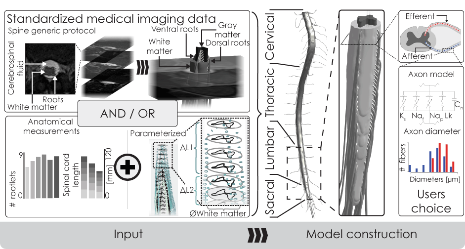
1. Anatomical Representation:
There is no consensus on which anatomical structures should be represented at different levels of resolution for various neural engineering treatments, and the underlying anatomical or histological data is often lacking. We are working to establish a validated standard for treatments that aim to modulate spinal circuits. Key contributions include accurately representing spinal rootlets with varying levels of detail, from simple tubular models to more anatomically precise spreads. Our findings have highlighted the importance of accurately modeling rootlet spread to avoid activation discrepancies, underscoring the need for anatomical precision in spine modeling.
2. Patient-Specific Modeling:
High-resolution imaging is crucial for capturing small-scale structures, such as the critically-important spinal rootlets, but current MRI protocols face challenges like lengthy imaging times, inconsistencies across machines, and resources that are not universally available across hospitals. We have started identifying and if necessary developing flexible and standardizable MRI acquisition strategies to address these limitations and improve data quality for patient-specific modeling.
3. 3D Volume Generation:
Generating precise 3D volumes is challenging due to the lack of standardized methods, often leading to labor-intensive and error-prone processes. We introduce parameterized routines that automatically assemble 2D anatomical outlines into 3D volumes representing macroscopic tissues, and fill these with streamlines representing microscopic structures. Additionally, we develop medical imaging segmentation and computer vision routines to extract 3D volumes from imaging data. We are currently working to make these computational tools broadly accessible to users with varying levels of expertise in modeling and image processing.
First Published on October 4, 2024
Last Updated on November 14, 2025 by Zhaoshun Hu